Reproductive System of Earthworm
Reproductive System of Earthworm
Earthworms are hermaphrodites but they cannot fertilize their own eggs because of their relative position of male and female genital aperture and they are protrandous (i.e. male sex mature earlier than female gametes). So, cross-fertilization takes place.
A] Male reproductive organs
They consist of following parts:
- Testes
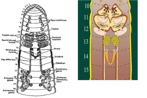
There are two pairs of small, white and lobed testes, located in 10th and 11th segment.
They lie ventro-laterally below the alimentary canal, close to mid-ventral line on either side of ventral nerve cord.
Each testis consists of 4-8 finger like projections/ processes, containing round cells called spermatogonia.
They are enclose within the testis sac.
Function: They produce sperm.
- Testis sac
Testes are enclosed by wide, thin-walled, whitish structure known as testis sac.
There are two testis sac present in 10th and 11th segment.
Testis sac communicates with a pair of seminal vesicles of succeeding segments.
The testis sac of 11th segment become larger as they enclosed the seminal vesicles of that segment.
They develop from the body cavity as small out-growth and lateral become saccular.
- Seminal vesicles
They are large, white, spherical structure found in two pairs, located in 11th and 12th segments.
They are also called septal pouches as they are formed as outgrowth of septa.
Seminal vesicles of 11th segment are smaller and are surrounded by testis sac of the same segment and communicates with the testis sac of 10th segment while those of 12th segment are free and communicate with the testis sac of 11th segment.
Function
Spermatogonia undergo maturation division to form spermatozoa or sperm.
Maturation of sperm takes place in seminal vesicles.
- Spermiducal funnel/ spermatic funnel
They are cup like curvature found in two pairs.
They are ciliated sperm funnels, lying below each testis in the segment 10th and 11th segment and enclosed within the same testis sac.
Function
Mature sperms from seminal vesicles move back to the testes sac and pass through the spermiducal funnel into vasa differentia.
- Vasa deferentia
They are elongated narrow ciliated thread like tubular structure which extends from 12th to 18th segment.
They are found in two pairs and each pair lie on either side of alimentary canal.
They extends from 12th to 18th segment.
In 18th segment they join together with a thick prostatic duct and forms common prostatic and spermatic duct.
Function
They collect sperm from spermatic funnel and give to prostate gland.
- Prostate gland (A gland in male that surrounds and opens into the urethra where it leaves the bladder)
There are a pair of large, white, flat, solid, irregular and lobulated gland, extending from 16th to 20th or 17th to 21st segment.
Each gland consists of large glandular part-containing many lobules and small non-glandular part-consisting of several small ductules.
From each prostate gland emerges a short, thick curved prostatic duct in 18th segment.
The prostatic duct joins the two vasa deferentia of its own side unite to form a common prostatic and spermatic duct.
Prostatic duct opens separately through a male genital aperture on the ventral side of 18th segment.
Thus each genital aperture has 3 separate apertures- two of the vasa deferentia and one of the prostatic gland.
Function
It produce prostatic fluid which is alkaline in nature. It activates sperms. And also it keeps sperm motile.
- Accessory glands
These are two pair of whitish, spherical structures found one pair in each of the 17th and 19th segment.
Found on the ventro-lateral body wall on either side of the ventral nerve cord.
They open to the exterior by a number of ducts on two pair of genital papillae, situated in 17th and 19th segment.
Secretion of these gland helps in holding two worms during copulation.
Function
They help in pseudocopulation.
- Male genital pore
It is found in one pair located in 18th segment.
Function
It acts as male genital pore.
Female reproductive organ
They includes following:
- Ovaries
A pair of small, whitish and lobulated ovaries located in the 13th segment, on either side of ventral nerve cord.
Each ovary consists of several finger like processes in which ova are arranged in a linear series in various stage of development (being mature in the distal part and immature in the proximal part).
Function: They form ova.
- Oviducal funnel
Below each ovary, there is a small saucer-shaped ovarian funnel with folded and ciliated margin, which leads into a short oviduct.
Function: Ova enter through oviducal funnel and travel backward along the oviduct.
- Oviduct
The two, short conical oviduct lies in 13th and 14th segment.
They run posteriorly and converge to meet below the nerve cord and open by a single median female genital aperture mid ventrally in 14th segment.
Function: They collect ova from ovary and give to female genital pore.
- Spermathecae
There are 4 pairs in each of 6th, 7th, 8th, 9th segment situated ventro laterally.
Each spermathecae is flask shaped consisting pear-shaped ampulla, short narrow neck and a short narrow elongated diverticulum.
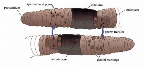
Spermathecae open outside through 4 pairs of spermathecal pores, situated ventrolaterally in the grooves between 5/6, 6/7, 7/8, 8/9 Segment.
Spermathecae are also called seminal receptacles as they store spermatozoa from another worm during copulation.
Function: They store sperm in diverticula during copulation and the ampulla provide nourishment for the sperm.
- Female genital pore
It is a single unpaired small pore, lies in 14th segment.
Copulation and cocoon formation
Copulation
The spermatozoa of one worm are transferred to another during a process called copulation.
OR, the process of transformation of sperms of one worm to another worm for the cross-fertilization is called copulation.
Copulation takes place during rainy season (from July to October) at night or early in the morning (dawn in the morning before sunrise).
During copulation two earthworms come closer and are ventrally attached in opposite direction in such a way that the male genital pore of one worm lie opposite to the spermathecal pore of other.
Both worms remain united together by the secretion of accessory glands and also by mutual penetration of setae in each other’s body.
Sperm and prostatic fluid of each worm are exchanged and released or stored in spermathecae through spermathecal pore.
Copulation lasts for about an hour.
Then the worm separate and later (after some day) they lay their eggs in cocoon.
Cocoon formation and Fertilization
Cocoon formation takes place after copulation, when ovaries mature.
The epidermis of clitellar region (14th, 15th 16th) contain 3 kinds of gland:
- Unicellular mucous gland- produce mucus for copulation
- Cocoon secreting gland- secrete wall of cocoon. Also secrete a gelatinous viscid and sticky substance which form a membrane girdle.
- Albumen gland cell- produce albumen; serves for nourishment of the growing embryo, which is deposited between the girdle and body wall.
The girdle soon harden on exposure to the air, into a tough but elastic tube which is called cocoon or egg capsule.
Then the worm starts to wriggle behind so that the girdle slipped forward. As the girdle pass over the female genital pore, it receives ova and when it passes over spermathecal, it receives sperm through spermathecal pores.
Finally, the girdle is thrown off from the anterior end and soon the elasticity of its wall closes up two ends to form a cocoon or ootheca.
Several cocoon are formed after each copulation because the spermatozoa in spermathecae do not pass out all at one time.
The cocoon are barrel shaped, light yellow in color and measuring about 2-2.5µm in length and 2.5-2µm in diameter.
Fertilization
Fertilization takes place inside the cocoon, where each ovum is fertilized by sperm.
As a rule, there is only one embryo develops (only one fertilized egg proceeds to develop new individual and rest degenerate). So the cocoon contains many fertilized eggs but only one earthworm can release from cocoon.
The development is direct i.e. no larval stage. The young worm when fully grown, crawls out of the cocoon in about 2-3 weeks which resembles the adult except in size and absence of clitellum.


Nice blog Mam
Quite impressive
This info is worth everyone’s attention. How can I find
out more?
Everything is very open with a clear description of the challenges.
It was truly informative. Your site is useful. Many thanks for sharing!
I think it is good but there is absence of three glands in clitellum.
Really useful. It’s clear and informative… Thank you for sharing
nyc notes..
Thank you so much
Very nice
Amazing mam 👏🙏🙇
Amazing mam🙇🙏👏
Its awesome
nice mam