
Detailed Structure of Frog’s Heart
Detailed Structure of Frog’s Heart
Heart of frog is three chambered. It is dark red colored conical muscular organ situated mid-ventrally in the anterior part of the body cavity in between two lungs.
The heart is enclosed in two membranes- an inner epicardium and outer pericardium.
The space between these two layers is called pericardial cavity. It is filled with pericardial fluid which performs
- Protects the heart from mechanical injury.
- Keeps the heart moist
- Allows the free movement during beating.
- Also keeps in keeping the heart suspended in its proper position.
External Structure of Heart
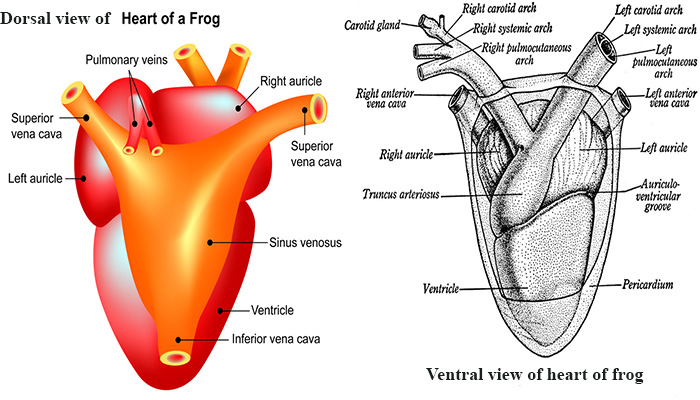
Externally heart looks like a triangular structure. It is reddish color.
It is 3 chambered besides sinus venosus and truncus arteriosus.
Its anterior end is broad and posterior end is somewhat pointed.
The anterior broarder part is called auricles whereas the posterior part is called ventricles.
Auricles are two chambered: left and right auricles. These auricles are demarcated externally by very faint longitudinal interauricular groove. So it externally appears one.
Ventricle is single chambered. It is most important part of the heart. It is conical in shape with thick muscular walls. It is clearly separated from auricles by coronary sulcus.
Heart of frog consists of two additional chambers:
- Sinus venosus- On the dorsal surface of heart, two precaval and a postcaval fused to form wide chamber called sinus venosus.
It is thin walled dark colored triangular structure which opens into the right auricle.
It receives impure blood from all parts of the body and pours it into right auricle.
- Truncus arteriosus
It is a tubular structure arise from the right side of the ventricle is called truncus arteriosus.
It extends forward over the right auricle and finally divides into two branches to form aortic trunks. Aortic trunk consists of- carotid arch, systemic arch and pulmonary arch.
Internal structure of heart of frog

The ventral view of internal structure of heart shows two auricles, one ventricle, truncus arteriosus and the valves to keep the blood flowing in one direction.
The wall of heart consists of three layers- outer epicardium, middle mesocardium and inner endocardium.
Internally heart is 3-chambered with two auricles and one ventricles.
The two auricles are separated from each other by interauricular septum. Right auricle is larger than left. In the right auricle, close to the septum there is a transverse oval opening called sinuauricular aperture. The blood enters into right auricles through this aperture. This aperture is guarded by two lip like flaps called sinu-auricular valves. These valves allow the flow of blood towards right auricles but prevent backward flow of blood.
In left auricle close to the septum there is a small opening of pulmonary vein which has no valves. The left auricles receives blood from lungs through pulmonary veins.
The ventricle has a thick muscular and spongy wall having numerous longitudinal clefts, separated from each other by muscular projections called columnae carnae. The two auricles open into a single ventricle chamber by auriculo-ventricular aperture which is guarded by two pairs of auriculoventricular valves. The valves are provided with chordae tendinae. The chordae tendinae pull the flaps backward to close the opening and thus prevent the valves from the backward flow of blood into the auricles.
From the right anterior side of the ventricle, truncus arteriosus arises. The ventricle opens into truncus arteriosus and the opening between these two is guarded by semilunar valves. When ventricles contract semilunar valves are pushed apart and make a free passage for the blood from the ventricles to truncus arteriosus but prevent back flow of blood from truncus arteriosus into the ventricles.
Internal structure of truncus arteriosus
The truncus arteriosus is divided into two unequal parts:
- Conus arteriosus or pylangium- the part near the ventricle- proximal part.
It is long broad and thick walled structure. It contains a large cavity called pylangium. It is provided with semilunar valves at its both ends.
Pylangium is incompletely divided into right cavum aorticum and left cavum pulmocutaneum by a spiral valve. - Bulbos arteriosus- the part away from the ventricle- distal part.
The synangium anteriorly divides into two branches. Each branch is called aortic trunk. Each aortic trunk is provided with three openings. They are carotid arch, systemic arch and pulmocutaneous arch.
Lovely I enjoyed it a lot very useful keep it up👍👍👍👍👍👍👍👍👍
I enjoyed this too much
Very good
Nice to study for students
Nicely Explained
Well understood by your text…short,sweet and clarifying
Thank u so much
Interesting
I LIKE THE WAY OF EXPLANATION. THANK YOU SO MUCH.
Amazing clarification …. Loved the way it it presented … Thanks a lot